NHRI Communications

會議報導
107年度國家衛生研究院生物醫學學術研討會紀實
Report on 2018 Biomedical Research Symposium of National Health Research Institutes
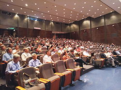 107年度國家衛生研究院生物醫學學術研討會(2018 Biomedical Research Symposium of NHRI)已於8月15日至16日假本院會議中心舉行。今年度會議依往例規劃2場特別演講(Plenary lecture)及6場專題演講,邀請國內外學者專家、執行成果優良之計畫主持人、本院研究人員及年輕研究學者,共26位講員口頭報告分享研究成果與經驗。本次會議參加人員除院外計畫學術審查之國內、外專家近五十餘位共同與會外,另有整合性計畫研究人員、本院研究人員及國內各機構研究人員等,總計近四百一十人次與會,可謂盛況空前,充分達到學術交流之目的。
本次會議邀請國立臺灣大學醫學院臨床醫學研究所陳培哲院士及中央研究院細胞與個體生物學研究所李奇鴻特聘研究員擔任大會特別演講講員。會議首日上午由本院梁賡義院長主持開幕,隨後由陳培哲院士以「Next Generation Challenges of Hepatitis B: From Novel Virology and Immunology to Virus Cure註1」為題發表演說;次日上午則由李奇鴻特聘研究員以「Antagonistic Regulation by Afferent-derived Factors to Ensure Appropriate Dendritic Field Sizes and Synaptic Partnership註2」為題發表演說。
而會議精心規劃的6場專題演講,主題分別為:「Biomedical Science」、「Population Health Research」、「Cancer Research」、「Medical Engineering」、「Molecular Genetics and Medicine」及「Neural Science」,共邀請國內外24位講員,於2個會場同步進行演講,分享最新的研究進展與寶貴的經驗,藉以讓與會者得到學術交流及研究合作的機會。
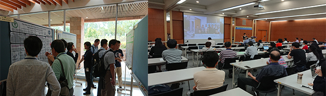
囿於演講場次有限,本次會議另安排二個壁報時段(Poster session),由整合性計畫主持人及本院研究人員張貼壁報展示研究成果(共計發表89篇壁報論文),並分組安排壁報論文討論會議(Poster discussion),由各組會議主持人邀請部分壁報展示之研究人員簡短報告分享研究成果,以促進壁報發表者、計畫主持人、所有分組與會人員及學術審查委員間之討論與互動,除有助於委員瞭解計畫執行成效,亦能協助解決研究過程可能遭遇之困難。
二天的學術活動中,藉由演講、壁報論文展示及分組討論會議等安排,引發許多熱烈的討論及腦力激盪,使與會人員獲益良多,且因交流互動增加,促成未來合作或團隊研究形成之可能,為國人之醫藥研究發展水準厚植潛力。
註1:陳培哲院士特別演講摘要(演講內容請見國衛院電子報第757期「影音節目」)
 Title: Next Generation Challenges of Hepatitis B: From Novel Virology and Immunology to Virus Cure
Abstract:
Hepatitis B control has been much improved in the last three decades, after universal vaccination and effective virus-suppression drugs implemented in the world. Despite of this impressive achievement, hepatitis B remains a main health issue and the world still harbors about 240 million hepatitis B carriers. Without treatment, about a quarter of them will eventually succumb to end-stage liver diseases, such as liver cirrhosis or hepatocellular carcinoma (HCC). Toward these unmet needs in hepatitis B, I try to review the new challenges and our recent research responses.
The most important unmet need is a HBV-curative therapy. Current antiviral regimens can only interrupt new cycle of viral replication but cannot eliminate the pre-existing viruses. Known HBV viral proteins or DNA molecules have been targeted to develop small molecules, in order to eliminate virus from infected hepatocytes. Despite of lot of efforts, the progress is still limited. We are interested in the biology of intriguing HBx protein, especially on its interaction with cellular proteins. By using model system, one cellular vesicle trafficking pathway may be shown involved with HBx function. We are in the process in genetic and functional identification of the exact viral molecule(s) as HBx targets. Moreover, we resumed the study of HBV spliced RNAs (discovered in Taiwan and Japan 30 years ago), and showed some of them required for effective HBV infection in human-hepatocyte chimera mice. If confirmed, these viral spliced RNAs and their encoded viral protein may reveal new HBV biology and become novel targets for antiviral development.
The second approach is to rejuvenate the exhausted anti-HBV immune system among the HBV carriers. Different from the exhausted immunity against cancer that can be reactivated by anti-PD1/PDL1 to combat human cancers, the same regimen failed to recover anti-HBV immunity to help HBV clearance. The immune exhaustion of T cells in chronic hepatitis likely takes different characters. In fact, HBV infection presented certain unique immune features. At first, acute HBV infection does not induce the conventional immune responses, such as transcriptional activations of IFN/IL1beta. Despite of this, the host still mount an effective adaptive immunity to clear HBV. We are interested in whether HBV infection induces a novel, unconventional innate immunity. The second feature is the immune clearance of HBV infection depends upon the age of infection. HBV infection in newborns always become chronic, but in the adults always become cleared. This age-dependent HBV immunity could be related to the gradual development gut microbiome after delivery, as shown in the HBV-mouse models. Such models allow us to dissect the hepatic cell population possibly participating these processes, and their working mechanisms.
The new challenges of HBV virology and immunology still wait to be resolved. Insights from the investigations may open up new avenues to develop better virus-curing therapies in the future..
註2:李奇鴻特聘研究員特別演講摘要(演講內容請見國衛院電子報第758期「影音節目」)
 Title: Antagonistic Regulation by Afferent-derived Factors to Ensure Appropriate Dendritic Field Sizes and Synaptic Partnership
Abstract:
During development, CNS neurons elaborate dendritic arbors in stereotypic patterns to synapse with appropriate partners. Dendritic patterning defects are found in many neuropsychiatric diseases, such as Autism spectrum disorders and Rett syndrome, and might underlie their cognitive deficits. However, little is known about how extracellular cues orchestrate dendritic development and how dendritic patterning defects disrupt neural circuit assembly and function. We use the first-order interneuron Dm8 in the Drosophila visual system as a model to tackle these questions. Each Dm8 neuron elaborates a large dendritic tree to receive synaptic inputs from ~14 R7 photoreceptors. In a forward genetic screen, we identified that insulin/Tor (Target of Rapamycin) signaling pathway positively regulates dendritic fields of Dm8s. Single Dm8 neurons devoid of insulin receptor, its substrate Chico or the downstream signaling components, Rheb and Tor, developed a smaller dendritic tree than wild-type did, while mutants lacking the negative regulators, Pten or Tsc1, had larger dendritic trees. Furthermore, the insulin-like peptide 2 (Dilp2) derived from lamina neurons, is delivered to the axonal terminals during development and is required for Dm8’s normal dendritic development. RNAi-mediated knockdown of Dilp2 in the lamina neuron L5, but not in photoreceptors, reduced the dendritic field size of Dm8s. Using a novel split-GFP tag system, we observed endogenous insulin receptors in developing dendrites of Dm8s. Thus, during development L5-derived Dilp2 activates insulin receptors on Dm8 dendrites to promote dendritic growth. We have previously shown that R7-derived Activin negatively regulates the sizes of Dm8 dendritic fields (Ting et al., 2014). However, mutant Dm8 devoid of both Activin and Tor signaling exhibited additive but highly variable dendritic phenotype, suggesting that these two signaling pathways operate in parallel.
Finally, using a novel activity-dependent receptor-based GRASP (GFP reconstitution across synaptic partners) method to probe active synaptic connections, we found that the enlarged dendritic field of Pten mutant Dm8 leads to aberrant connections with neighboring R7s while Tor mutant Dm8 receives few if any active synaptic input from R7s. In summary, the size of Dm8’s dendritic field is controlled by two parallel signaling pathways operating in opposite directions: Dilp2 derived from non-synaptic partner lamina neurons positively regulates Dm8’s dendritic field sizes, while Activin from Dm8’s synaptic partner R7s negatively regulates Dm8. We argue that the antagonistic regulation of dendritic growth ensures robust size-control of Dm8’s dendritic fields.
《文/圖:學術發展處蕭振祥》